New clues about human cardiovascular evolution
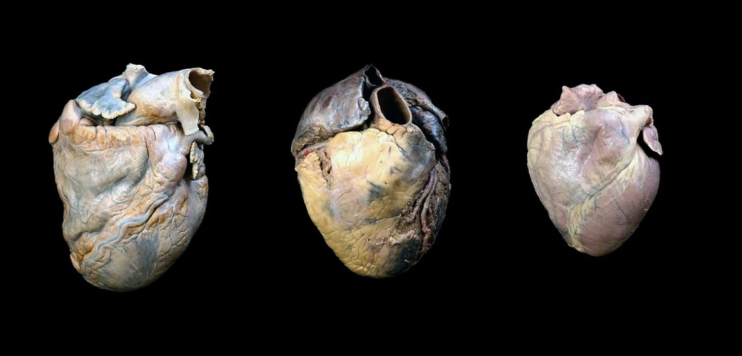
Studies of the aorta and the heart anatomy of humans and living great apes show some important differences.

There's hardly a system more important to life than the heart and circulatory system. Every schoolkid learns the basics of circulatory system anatomy and function.
Hearts like ours have four chambers. The right atrium receives deoxygenated blood from the major veins of the body, the inferior vena cava and superior vena cava. When the atria contract, this deoxygenated blood passes into the right ventricle, which sends it on its way to the lungs via the pulmonary arteries. Oxygenated blood returns from the lungs into the left atrium, then into the left ventricle, and then pumped by ventricular contraction into the body through the aorta. The four-chambered structure of the heart keeps oxygenated and deoxygenated blood separate in a continuous circuit. That structure is conserved across all living mammals.
Still, mammal hearts are not all alike. They differ in heart shape and size, in heartbeat pace and cadence, and in countless smaller anatomical details.
In some of these details, humans stand out from chimpanzees and other living great apes. The challenge is understanding why these differences evolved. Some fascinating papers over the past few years have examined differences in cardiovascular anatomy between humans and living great apes.
Aortic root
The aorta is the artery that carries oxygenated blood from the left ventricle out to the body. It leaves the heart going upward and quickly arches downward, branching off the arteries of the arms and neck on its way. In 2023, Luis Ríos and coworkers published the results of a study of the aorta of humans, chimpanzees, and gorillas.
The study included data taken by echocardiography from more than 900 humans, 88 captive chimpanzees, and 65 captive western gorillas. The idea was to understand how the aorta is scaled to body mass in these primates.
Ríos and collaborators mainly focused on one measurement: the diameter of the aortic root. The aortic root is the very start of the aorta that supports the flaps of the aortic valve. In humans this is generally around 25 to 40 mm in diameter. An enlarged aortic root much beyond this range is a considered an aneurism, which may have a risk of rupture or separation of the aortic wall, a catastrophic injury known as an aortic dissection. Measurement of the diameter of the aortic root is a standard diagnostic procedure during examination of the heart both in human and veterinary practice.
Gorillas average larger hearts and larger aortic measurements than humans. That's no surprise: bigger bodies, bigger hearts, bigger aorta. There is a good bit of overlap between the size distributions of humans and gorillas, and that parallels the distribution of body mass.
But Ríos and collaborators found that the aortic root scales differently than body mass between these species. Gorillas average a lot higher body mass than the human sample, yet only a little higher in aortic root diameter. Comparing aortic root to body mass, humans are disproportionately large.

Ríos and coworkers suggest that the comparatively larger size of human aortas may reflect the higher energy expenditure of humans compared to the other living great apes.
The descending aorta
After the aorta leaves the heart, it curves downward inside the lower thorax and abdomen. This portion, called the descending aorta, runs close against the front surfaces of the vertebrae and intervertebral discs.
In humans the descending aorta often creates a flat impression on the front surfaces of the vertebral bodies. The aorta runs down slightly to the left of center, and that means that the flattened surface is on the left. The result is a vertebral body that is slightly out of round, asymmetrical when viewed from above or below.
Ríos and collaborators measured this asymmetry in the skeletons of 48 adult humans, as well as 43 chimpanzees, 24 gorillas, and 6 orangutans. They confirmed the asymmetrical shape of human vertebrae, most prominently across the lower throracic spine from the sixth to twelfth thoracic vertebrae. The curved path inscribed by the descending aorta is clearly visible in the wave of asymmetry moving down the spine. The great apes have much less asymmetry, across a smaller range of vertebrae than in humans.
Why do many humans have such a marked impression for the descending aorta? The aorta’s size itself may not be too important. A gorilla’s aorta is equally large, after all. It may be a combination of factors. In humans, the flatter thorax puts a premium on space from front to back. Our upright posture puts different stresses on the aorta-vertebral interface.

Having a trait that can be observed on bones means that the research could include fossils! The team roped in a small sample: the Turkana Boy vertebral skeleton attributed to Homo erectus, as well as one vertebra from a Neanderthal from El Sidrón, Spain. They found that the Neanderthal has flattened area on the left similar to most modern people, while the Turkana Boy vertebrae had little or no evidence of asymmetry.
It’s not hard to improve on that fossil sample. I took a quick look at other fossils: Homo erectus vertebrae from Dmanisi, Georgia, vertebrae of Homo naledi from the Rising Star cave system, the StW 431 and Sts 14 vertebrae attributed to Australopithecus africanus. One or two of the lower thoracic vertebrae from Rising Star do have what may be a flattening on the left side. However these specific fossils have some erosion on their front surfaces and I’d like to do a more careful study to see if the asymmetry might have resulted from postmortem damage. The others don’t have any evident flattening or asymmetry that I would attribute to the aorta.
Ríos and coworkers suggested several possibilities for why humans and Neanderthals might be different from Homo erectus. These ideas centered on lower cardiovascular needs for early Homo.
That idea might have something to it. Still, I think that a fuller comparison with humans of varied body sizes and across a wider range of developmental ages would be valuable. Modern people and Neanderthals have quite large vertebral bodies compared to many earlier hominins, and it may be that vertebral size or protrusion into the abdomen are the main influences on the asymmetry.
The ventricular twist
In a study that came out last year, Bryony Curry and coworkers looked at a different area of the heart: the apex of the left ventricle. The left ventricle is the most powerful of the heart’s chambers, and its apex plays a key role increasing this power.
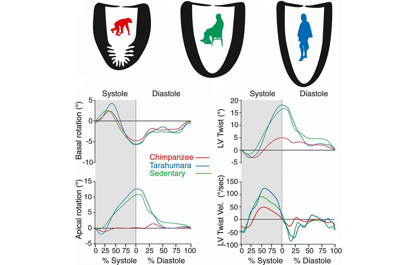
The ventricle doesn't just contract like a simple balloon but instead carries out a twisting motion. The base of the heart rotates in one direction, while the apex rotates in the opposite direction. That twisting motion has the effect of wringing out the maximum volume with each heartbeat.

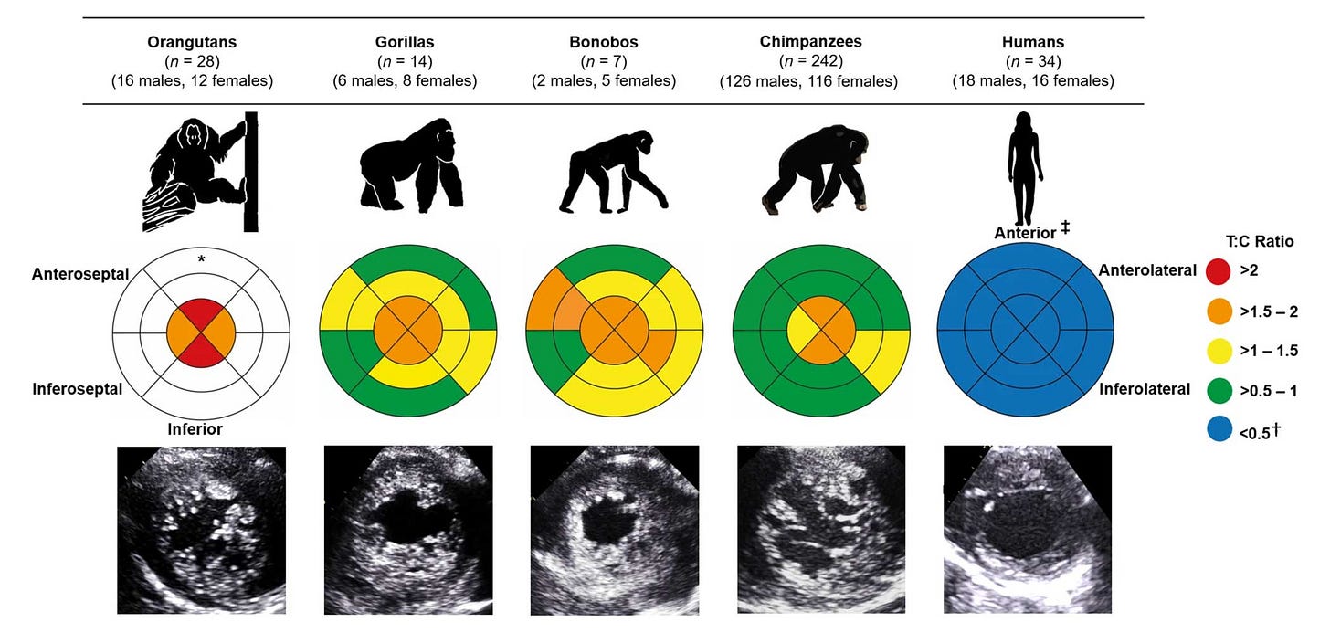
Curry and coworkers used echocardiography to examine the left ventricles of chimpanzees, bonobos, gorillas, and orangutans. These primates tend to have a greater development of the sponge-like network of muscle fibers and deep recesses within the left ventricle, known as ventricular trabeculation.
Trabeculations are a normal part of heart development and are present in every human heart. But the apex of the left ventricle typically has little trabeculation, which may relate to its function in the twisting motion during ventricular contraction.
In 2019, Robert Shave and collaborators examined the performance of the left ventricle in chimpanzees, sedentary people from the U.S., and people from the Tarahumara ethnic group in Mexico. Their research started from a focus on whether human cardiovascular system is better at endurance efforts.
Tarahumara people are subsistence farmers, with a high level of physical work most days. They have attracted a lot of attention from anthropologists and exercise physiologists because some of their traditions center on long-distance running. These people manifest a somewhat elongated left ventricle and more pronounced twisting motion when it contracts.

Chimpanzees are very different from humans in the low degree that their left ventricle twists during contraction. The 2024 work by Curry and colleagues confirmed that bonobos, orangutans, and gorillas also have this low twist. The results from these other apes show that humans are the odd ones out, with high twist and low trabeculation.
It’s not clear what the functional importance of the trabeculation itself may be for these primates. Within humans, cardiac muscle varies a great deal in its trabeculation. Both high and low trabeculation can be associated with cardiac disease. The trabeculation adds complexity and resilience to the heart tissue’s structure, and may be important in the maintenance of heart function across the lifespan.
Adapted for endurance?
The comparative anatomy of these cardiovascular traits helps reveal some of the demands on the human circulatory system. Shave and coworkers suggested that endurance performance might explain the difference between human and great ape hearts.
In their description, chimpanzee and gorilla activity patterns tend to have short bursts of high-intensity movement interspersed among long periods of low to moderate physical exertion. Humans in traditional societies have some activities that require high exertion over a longer period of time, endurance. Such endurance activities require a consistent high cardiac output.
Endurance and hard work are increasingly recognized as important to the collection of wild foods in hunting and gathering societies. One extreme version of this idea is the so-called “endurance hunting” hypothesis: the idea that ancient humans typically ran down their prey in day-long chases. But we don’t need to imagine ancient marathon hunts to recognize that human subsistence requires some long, hard work.
A lot of what scientists know about human evolution comes from fossils of ancient human relatives. This record of bones and teeth of ancient species leaves a lot of their bodies that we don't have direct evidence about.
New research is expanding the data on the anatomy of the heart and circulatory system in living humans and nonhuman primates by taking advantage of 3D imaging modalities. CT scanning and ultrasound on living nonhuman primates, mostly captive animals in zoos or sanctuaries, has become an important source of information about the anatomical structure and variation of our closest relatives.
Together with research on the cardiovascular performance of humans across societies, I expect we’ll learn quite a lot more about the hearts of our ancestors evolved.
Notes: The chapters by Scott Williams and Marc Meyer provide great overviews of the vertebral morphology of Australopithecus and early Homo.
References
Curry, B. A., Drane, A. L., Atencia, R., Feltrer, Y., Calvi, T., Milnes, E. L., Moittié, S., Weigold, A., Knauf-Witzens, T., Sawung Kusuma, A., Howatson, G., Palmer, C., Stembridge, M. R., Gorzynski, J. E., Eves, N. D., Dawkins, T. G., & Shave, R. E. (2024). Left ventricular trabeculation in Hominidae: Divergence of the human cardiac phenotype. Communications Biology, 7(1), 1–9. https://doi.org/10.1038/s42003-024-06280-9
Kraft, T. S., Venkataraman, V. V., Wallace, I. J., Crittenden, A. N., Holowka, N. B., Stieglitz, J., Harris, J., Raichlen, D. A., Wood, B., Gurven, M., & Pontzer, H. (2021). The energetics of uniquely human subsistence strategies. Science, 374(6575), eabf0130. https://doi.org/10.1126/science.abf0130
McGurk, K. A., Qiao, M., Zheng, S. L., Sau, A., Henry, A., Ribeiro, A. L. P., Ribeiro, A. H., Ng, F. S., Lumbers, R. T., Bai, W., Ware, J. S., & O’Regan, D. P. (2024). Genetic and phenotypic architecture of human myocardial trabeculation. Nature Cardiovascular Research, 3(12), 1503–1515. https://doi.org/10.1038/s44161-024-00564-3
Meyer, Marc R. & Williams, Scott A. (2019). The spine of Early Pleistocene Homo. In Been, Ella, Gómez-Olivencia, Asier, & Kramer, Patricia A. (Eds.), Spinal Evolution: Morphology, Function, and Pathology of the Spine in Hominoid Evolution (pp. 153–183). Springer. https://doi.org/10.1007/978-3-030-19349-2_8
Moittié, S., Baiker, K., Strong, V., Cousins, E., White, K., Liptovszky, M., Redrobe, S., Alibhai, A., Sturrock, C. J., & Rutland, C. S. (2020). Discovery of os cordis in the cardiac skeleton of chimpanzees (Pan troglodytes). Scientific Reports, 10(1), 9417. https://doi.org/10.1038/s41598-020-66345-7
Ríos, L., Sleeper, M. M., Danforth, M. D., Murphy, H. W., Kutinsky, I., Rosas, A., Bastir, M., Gómez-Cambronero, J., Sanjurjo, R., Campens, L., Rider, O., & Pastor, F. (2023). The aorta in humans and African great apes, and cardiac output and metabolic levels in human evolution. Scientific Reports, 13(1), 6841. https://doi.org/10.1038/s41598-023-33675-1
Shave, R. E., Lieberman, D. E., Drane, A. L., Brown, M. G., Batterham, A. M., Worthington, S., Atencia, R., Feltrer, Y., Neary, J., Weiner, R. B., Wasfy, M. M., & Baggish, A. L. (2019). Selection of endurance capabilities and the trade-off between pressure and volume in the evolution of the human heart. Proceedings of the National Academy of Sciences, 116(40), 19905–19910. https://doi.org/10.1073/pnas.1906902116
Williams, Scott A. & Meyer, Marc R. (2019). The spine of Australopithecus. In Been, Ella, Gómez-Olivencia, Asier, & Kramer, Patricia A. (Eds.), Spinal Evolution: Morphology, Function, and Pathology of the Spine in Hominoid Evolution (pp. 125–151). Springer. https://doi.org/10.1007/978-3-030-19349-2_7

